Preoperative navigated transcranial magnetic stimulation (nTMS)
The navigated transcranial magnetic stimulation (nTMS) is applied preoperatively for the localization of important brain regions, such as the speech or motor area. The physical basis for this is an activation or suspension of nerve cells by using magnetic pulses.
Such a navigated (= deliberately provoked by the computer) impulse on the surface of the brain induces on the motor cortex (movement area) the activation of neurons, which control muscles (e.g. arm and leg muscles). Thus, motor evoked potentials (MEPs) of defined muscles can be derived if the navigated pulse exactly stimulates the corresponding motor cortex area.
Repeated magnetic stimulation leads to temporary functional suspension of nerve cells. This principle is utilized for the identification of relevant language areas. The patient is presented with various graphics on a monitor, which are to be determined/named. In the first stage the goal is to find out, which presented pictures the patient may name clearly and repeatedly securely (positive images). Subsequently, the positive images are presented again and simultaneously a navigated, repetitive magnetic stimulation of the cerebral hemisphere is performed. In this way, in certain cortical areas language production errors, phonetic errors, semantic errors and non-designation are provoked, which can be identified as possible language-related areas. In addition, the examiner also learns that those stimulated areas, which provoke no speech impediment, are not language-related areas.
As the magnet stimulation is performed by navigation, cortical areas where either MEPs (motor cortex mapping) or speech impediment are evoked (language mapping) can be accurately localized and can be color-coded in the MRI data set.
Procedure:
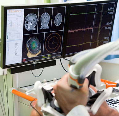
1. For the navigated transcranial magnetic stimulation the device from the company Nexstim® is used. The examination takes place in a quiet room so that the patient and the examiner can concentrate entirely on the examination.
2. First, the MRI data set of the patient is imported to the computer.
3. In order to achieve synchronization of the patient with the MRI data set, the computer must be - with the help of a special display instrument - shown where certain fixed points of the patient are located. A camera recognizes both the patient's head (via a headband) and the points shown by the examiner (via the pointing device).
4. For recording muscle activation (motor evoked potentials; MEPs) recording electrodes are glued to the muscles, which are to be examined. These continuously register the muscle activity (here the hand muscles).

5. The coil of transcranial magnetic stimulation is also recognized by the computer. Therefore, a navigated stimulation of defined cortical areas can be carried out. The activation of the hand muscles then shows potential changes in the field of hand-MEPs. Those areas where stimulation has been successful (triggered MEPs) are highlighted. Thus, at the end of the examination MRI data set is obtained, in which the motor cortex area is highlighted in colour. We can use this for our navigation system during the operation, thereby avoiding damage to movement areas.