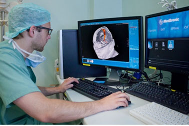
Preoperative diffusion tensor imaging Fibre Tracking (tractography)
The "diffusion tensor imaging" - magnetic resonance tomography (DTI-MRI) is a special sequence of magnetic resonance imaging. In this the movement (diffusion) of hydrogen particles in three-dimensional alignment along the nerve fiber extensions is registered. With special software it is now possible to display the detected motions as "tensors" and to connect them with each other. In this way, fiber pathways can be identified in the brain and then color-coded in the magnetic resonance data set. The fiber pathways most commonly include the pyramidal tract and the visual pathway.
Procedure:
- The DTI-MRI data is processed using special software on the surgical planning station.
- A starting and end point of the fiber webs are specified for the software. The software then determines in which three-dimensional direction, the "tensors" are most aligned (the largest vector) and then connects these points from the starting to end point. For exact location of the starting point in the movement area the identified by NTMs-regions can be chosen. Thus, both examinations complement each other and ensure a highly accurate preoperative functional model of the patient.
After depiction of the desired fiber webs, relations of lesions to eloquent areas may be estimated on the virtual model. This enables an optimal surgical preparation.


