
Infantile Hydrocephalus
The causes of hydrocephalus in children are diverse. It can be caused, for example by bleeding in the brain water chambers after birth, especially in preterm infants, by malformations of the spinal cord, caused by infections of the meninges, the cerebrospinal fluid or the brain, by traumatic brain injury or tumors. In addition, it can also occur within the scope of genetic syndromes with malformations of the brain.
It is characterized by a blockage of the cerebrospinal fluid chambers which are located within the brain. If the cerebrospinal fluid chambers become too large, vital parts of the brain are compressed, especially when the skull is already tightly closed.
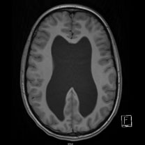
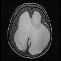
Depiction of the distinctly enlarged cerebral fluid chambers in two different cases of infant hydrocephalus in MRI imaging (left T1- and right T2-weighted)
The hydrocephalus becomes visible in young children with open fontanelle usually by an increased head growth. This means that the head is very big and the fontanelle is stretched thickly. But also slightly older children (2-6 years) may show an increased head growth, yet often accompanied by earlier clinical symptoms of an accumulation of cerebrospinal fluid such as increased fatigue, vomiting, blurred vision and gait disturbances. These symptoms are observed by the pediatrician and parents.
Further diagnosis in case of children with not yet closed fontanelle is done via ultrasound, which already depicts the enlarged cerebrospinal fluid chambers. This is often followed by a magnetic resonance imaging of the brain in order to clarify the exact cause. In the case of a closed cranium a MRI is directly performed.
If the cause of hydrocephalus (for example a tumor) can be removed, this is the treatment of choice. For other causes of brain water chamber widening two treatment methods come into question, whereby it should be defined individually which option is the most favorable for the respective patient. Either the connection of the cerebrospinal fluid chamber to the basal cistern by means of an endoscope (ETV) without implantation of a foreign object or in the other case the insertion of a shuntto ensure permanent brain drainage.

