
Endoscopic third ventriculostomy (ETV)
Our clinic also offers various endoscopic techniques in the context of treatment for hydrocephalus, some of which are carried out by only a few centers worldwide. An established technique is the Endoscopic third ventriculostomy (ETV) which can be a treatment option for certain forms of hydrocephalus and can avoid shunt placements. If a relocation of cerebrospinal fluid circulation is caused, for example, by a narrowing between the 3rd and 4th cerebrospinal fluid chambers (aqueduct), a so-called occlusive hydrocephalus can occur. Tumors within the cerebrospinal fluid chambers can move the conveyance routes and lead to an occlusive hydrocephalus. In these forms of hydrocephalus through an ETV a connection between the 3rdventricle and the outer CSF spaces can be created. Through a small hole in the skull bone the endoscope is introduced into the 3rd ventricle and its bottom is fenestrated by a perforation on the outer CSF spaces (basal cisterns). This allows an unobstructed CSF flow, made possible by bypassing the obstacle.
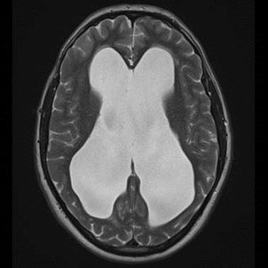
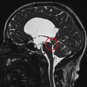
MRI images of occlusive hydrocephalus caused through narrowing by the link between the 3rd and 4th ventricle (aqueduct). Left: Depiction of the clearly extended lateral ventricle. Right: view of the aqueductal stenosis.
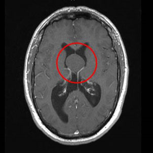
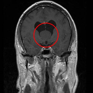
MRI images showing an occlusive hydrocephalus due to tumor in the 3rd ventricle.
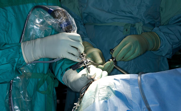
Performance of ETV in the operating room

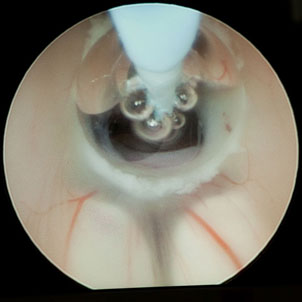
Intraoperative endoscopic view during an ETV. Left: Fenestration of the bottom of the 3rd ventricle to the external CSF spaces by coagulation. Right: enlargement of the opening to the external CSF spaces by means of a balloon catheter.

