
Image-guided radiotherapy (IGRT)
With modern radiotherapy techniques it is possible to create highly complex three-dimensional dose distributions closely conformal to the target volume while sparing neighbouring organs at risk. A prerequisite is the exact and stable positioning of the patient during treatment, so that the steep dose gradients around the target volume will not cause under-dosage of the target or over-dosage of critical structures. To achieve this, modern linear accelerators are equipped with integrated imaging systems, so that possible deviations from the planned patient set-up can be assessed and corrected before the start of each treatment fraction.
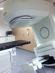
At the Department of Radiotherapy and Radiation Oncology, three different linac-based imaging techniques are available: the treatment beam line (TBL) operating at 6 MV, a dedicated "Image beam line" (IBL) of nominally 1 MV, and an X-ray tube of 70-121 kV mounted at one linear accelerator (kVision, see figure).
All three imaging systems have been physically and dosimetrically characterised and commissioned in the treatment planning system. The Siemens Artiste linear accelerator is thus completely modelled in the treatment planning system including the kV-CBCT technique, so that the imaging dose can be individually calculated and included in the treatment plan for each patient.
Publications about IGRT:
- Dzierma Y, Nuesken FG, Licht NP, Ruebe C. Dosimetric properties and commissioning of cone-beam CT image beam line with a carbon target. Strahlenther Onkol. 2013 Jul;189 (7):566-72. doi: 10.1007/s00066-013-0330-5. Epub 2013 May 30. PubMed PMID: 23715886.
- Dzierma Y, Nuesken F, Otto W, Alaei P, Licht N, Rübe C. Dosimetry of an in-line kilovoltage imaging system and implementation in treatment planning. Int J Radiat Oncol Biol Phys. 2014 Mar 15;88(4):913-9. doi:10.1016/j.ijrobp.2013.12.007. Epub 2014 Jan 20. PubMed PMID: 24456996.
- Dzierma Y, Ames E, Nuesken F, Palm J, Licht N, Rübe Ch. Image quality and dose distributions of three linac-based imaging modalities. Strahlenther Onkol. 2015; 191(4):365-374.

